
No Extraction Needed: How We Saved a Lower Molar with Root Canal & E-max Onlay
Age : 45-year-old
Gender : Female
Chief Complaint : “Severe pain in the lower left molar, especially when chewing.”
Clinical & Radiographic Findings
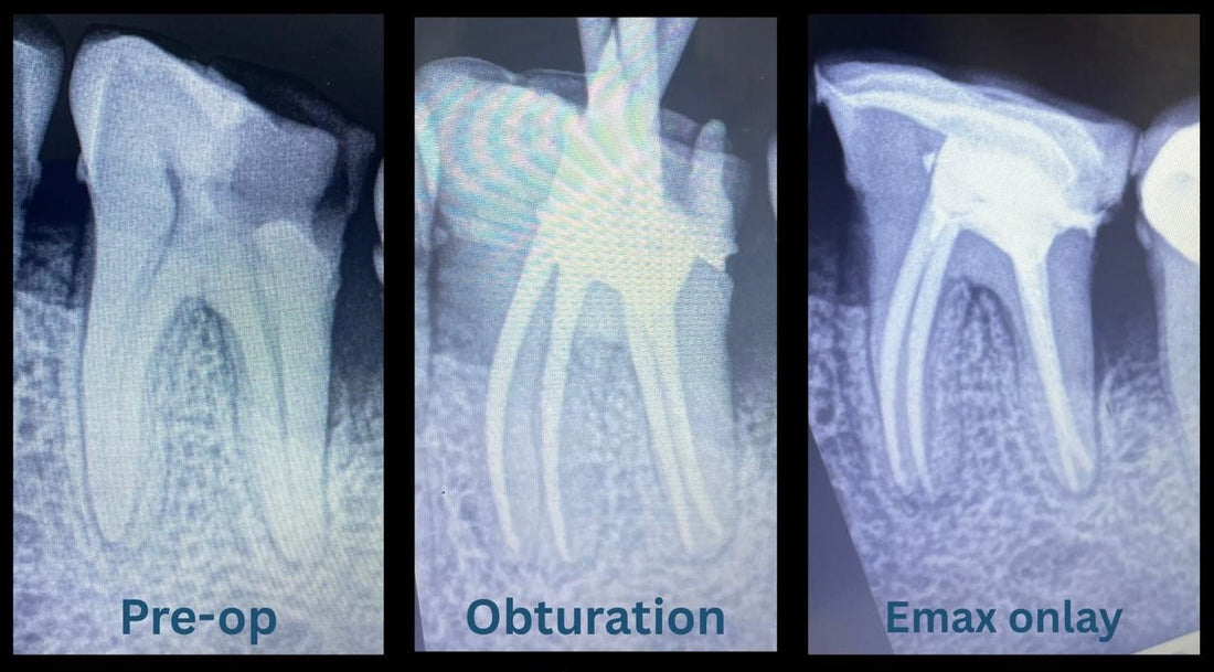
Tooth Involved: Mandibular left first molar
Clinical Examination: Tooth was tender to percussion with a deep carious lesion extending into the pulp.
Radiographic Assessment: Radiolucency reaching the pulp chamber causing irreversible damage to pulp
Treatment Protocol
1. Endodontic Treatment (RCT)
Access & Cleaning: Access cavity prepared; canals located and instrumented using rotary NiTi files with copious irrigation (NaOCl, EDTA).
Obturation: Canals filled with gutta-percha coated with bio ceramic sealer; postoperative radiograph confirmed adequate fill.
2. Post-Endodontic Restoration with E-max Onlay
Preparation: After RCT, significant tooth structure was lost occlusally. Instead of a full crown, motivation was to preserve maximum tooth structure with an Onlay.
Impression & CAD-CAM Workflow: Tooth prepared, impression taken, and an E-max ceramic Onlay fabricated in the dental laboratory. The Onlay was tried in, with occlusion and fit verified.
Cementation: Cemented with dual-cure resin cement after appropriate adhesive protocols ensuring isolation and clean margins
Outcome
Immediate Result: Patient reported complete resolution of pain upon biting and percussion.
Follow-Up: Re-evaluation at one week confirmed the tooth was asymptomatic with intact occlusion and restoration.
Long-Term: No complications or sensitivity reported at subsequent check-ups.
